44 labels of the human brain
Free Nervous System Worksheets and Printables Human Brain Clipart and Printables - You'll find a great collection of clipart and printables showing the human brain and nerve cells. Neuron Printable Clipart and Labeling Sheets - This is an amazing selection of neuron, or nerve cell, clip art for you and your kids to print and label. Climate changed the size of our bodies and, to some extent ... The researchers say there is good evidence that human body and brain size continue to evolve. The human physique is still adapting to different temperatures, with on average larger-bodied people ...
Labeled imaging anatomy cases | Radiology Reference ... URL of Article. This article lists a series of labeled imaging anatomy cases by body region and modality. On this page: Article: Brain. Head and neck. Spine. Chest. Abdomen and pelvis.

Labels of the human brain
Label-free imaging of human brain tissue at subcellular ... The human neocortex is conventionally divided into six layers [ 28 ]. These layers - molecular, outer granular, outer pyramidal, inner granular, inner pyramidal, and pleomorphic layers - are roughly distinguishable in MPM data ( Figure 2 A; representative higher-magnification images from each layer are shown in Figure S3 ). Draw And Label The Human Brain Structure - Draw Neat ... The brain structure is composed of three main parts: The structure and function of the human brain. The skull is composed of two sets of bones cranial and facial. Brain diagram, pituitary gland, anatomy, adhd, draw, search, to draw. How to draw a the human brain easy and step by step. 14 Informative Facts, Diagram & Parts Of Human Brain For Kids Human brain and the spinal cord are covered by three layers of tissue collectively known as the meninges. A clear fluid called cerebrospinal fluid flows between these layers (10) (11). The left part of the brain is responsible for analytical thinking. The right part helps in creative thinking (12).
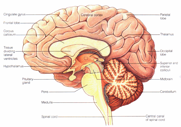
Labels of the human brain. Brain: Atlas of human anatomy with MRI - e-Anatomy - IMAIOS Choroid plexus of fourth ventricle Choroid plexus of lateral ventricle Choroid plexus of third ventricle Choroidal fissure Cingulate gyrus Cingulate sulcus Cingulum Circular sulcus of insula Cistern of lamina terminalis Cistern of lateral cerebral fossa Claustrum Collateral eminence Collateral sulcus Left Brain vs. Right Brain: Characteristics Chart ... Brain dominance theory is absorbing and enjoyable, plus it allows people to think about stereotypes and labels. In reality, though, psychology is complicated, and the truth is that there are very few people who have the traits of only one of these descriptions. How Well Do You Know About Parts Of The Brain? - ProProfs 1. What is the biggest part of the brain? A. Cerebrum B. Thalamus C. Brain stem D. Cerebellum 2. The cerebrum controls how we ____________. 3. What is the back part of the brain called? A. Thalamus B. Cerebellum C. Cerebrum D. Brain stem 4. Name one thing your cerebellum controls _________________. 5. Third harmonic generation imaging for fast, label-free ... (D) Infrared photons (white arrow) are focused deep in the brain tissue, converted to THG (green) and SHG (red) photons, scattered back (green/red arrows) and epi-detected. The nonlinear optical processes result in label-free contrast images with sub-cellular resolution and intrinsic depth sectioning.
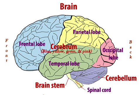
Brain charts An international team of researchers has created a series of brain charts spanning our entire lifespan - from a 15 week old fetus to 100 year old adult - that show how our brains expand rapidly in early life and slowly shrink as we age.. The charts are the result of a research project spanning six continents and bringing together possibly the largest ever MRI datasets ever aggregated ... Positions and Functions of the Four Brain Lobes | MD ... Brain Lobes and their Functions The brain is divided into four sections, known as lobes (as shown in the image). The frontal lobe, occipital lobe, parietal lobe, and temporal lobe have different locations and functions that support the responses and actions of the human body. Let's start by identifying where each lobe is positioned in the brain. Eight ways scientists are unwrapping the mysteries of the ... Mapping these connections isn't easy—there may be as many as 100 trillion connections in the human brain, and they're all tiny. Researchers need to find the best ways to label specific ... When makes you unique: Temporality of the human brain ... to figure out what makes a human brain identifiable at the level of functional neuroimaging correlates, we explored the temporality of brain fingerprints along two complementary directions: (i) what is the optimal time scale at which fingerprints integrate sufficient information and (ii) when does better identification of a fluctuating pattern …
The human brain: Parts, function, diagram, and more The brain is the human body's control system, and is part of the central nervous system (CNS). It connects to the spine and controls personality, movement, breathing, and other crucial processes ... Structures Of The Brain Quiz! Ultimate Trivia - ProProfs Structures Of The Brain Quiz! Ultimate Trivia. . 1. This structure controls thought, voluntary movement, language, reasoning and perception. 2. This is a collection of axons that connect the right and left hemispheres of the brain. 3. This part controls vision, hearing, eye movement, and body movement. Cross-sectional anatomy of the brain - e-Anatomy - IMAIOS Quadrigeminal cistern; Cistern of great cerebral vein Red nucleus Rhinal sulcus Rostrum of corpus callosum - Corpus callosum Septum pellucidum Short gyri of insula Sigmoid sinus Simple lobule [H VI and VI] Sphenoparietal sinus Spinal cord Splenium of corpus callosum - Corpus callosum Straight gyrus Straight sinus Parts of the Brain Activity for Kids, Brain Diagram, and ... PARIETAL LOBES - The parietal lobe provides sensory information to the brain including touch, pain and temperature. OCCIPITAL LOBES - The occipital lobe processes and interprets everything we see TEMPORAL LOBES - The temporal lobe controls emotions and short-term memory
Sagittal View of Brain Quiz
Parts of the brain: Learn with diagrams and quizzes | Kenhub Labeled brain diagram First up, have a look at the labeled brain structures on the image below. Try to memorize the name and location of each structure, then proceed to test yourself with the blank brain diagram provided below. Labeled diagram showing the main parts of the brain Blank brain diagram (free download!)

TDP-43 mutant transgenic mice develop features of ALS and frontotemporal lobar degeneration | PNAS
Brain: Anatomy, Pictures, Functions, and Conditions The cerebral cortex is the part of the brain that makes human beings unique. Functions that originate in the cerebral cortex include: Consciousness Higher-order thinking Imagination Information processing Language Memory Perception Reasoning Sensation Voluntary physical action 1 The cerebral cortex is what we see when we look at the brain.

Flashcards - Chapter 12 CNS - Anatomy of the Brain THE BRAIN embryonic development | StudyBlue
Human Brain: Facts, Functions & Anatomy | Live Science The human brain is divided into two hemispheres, the left and right, connected by a bundle of nerve fibers called the corpus callosum. The hemispheres are strongly, though not entirely,...
A.I. in the Ditch – Science, POLITICS, & Religion
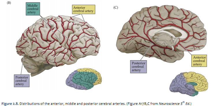
Lateral view of the brain: Anatomy and functions - Kenhub The lateral view of the brain shows the three major parts of the brain: cerebrum, cerebellum and brainstem . A lateral view of the cerebrum is the best perspective to appreciate the lobes of the hemispheres. Each hemisphere is conventionally divided into six lobes, but only four of them are visible from this lateral perspective.

TooSogiE Medical Images: Cranial Nerves : I - V
3D molecular phenotyping of cleared human brain tissues ... Although SHIELD 25 was fully applied to clear formalin-fixed 2-mm-thick coronal blocks of the human brain (9 × 5.5 × 0.2 cm 3) and label with different markers, our replication of this approach ...

brain-anatomy - The Mind Voyager
Ventricles of the Brain Function, Anatomy & Diagram ... The brain is a very fragile organ within the human body that relies on a lot of energy in the form of glucose. For that reason, CSF is highly important for the brain to survive and thrive ...
34 Label And Color The 3 Major Parts Of The Brain - Best Labels Ideas 2020
A unified 3D map of microscopic architecture and MRI of ... In each specimen, consecutive sections located rostral and caudal to the corpus callosum were processed for Nissl (thionin, to label glial and neuronal cell bodies), silver staining (Bielschowsky for labeling nerve fibers), and parvalbumin staining (immunolabeling of interneurons).
Human Anatomy for health & wellness Unit Plan
Parts of the Human Brain | Anatomy & Function - Video ... The parts of the brain include the cerebrum, the cerebellum, the brain stem, and the pituitary gland. The brain structure is protected by the skull, which is composed of the cranium and the bones...

WMU Psychology Department: Lisa Baker
Anatomy of the Brain | Simply Psychology Anatomy of the Brain. The brain receives information from sensory receptors and sends messages to muscles and glands. It is the centre of all conscious awareness and is divided into different lobes with different functions. It contains the cerebrum which makes up about 85% of the total mass.

35 Label The Brain Anatomy Diagram
Human brain - Wikipedia The brainstem includes the midbrain, the pons, and the medulla oblongata. Behind the brainstem is the cerebellum ( Latin: little brain ). The cerebrum, brainstem, cerebellum, and spinal cord are covered by three membranes called meninges. The membranes are the tough dura mater; the middle arachnoid mater and the more delicate inner pia mater.

Pin on The Brain and Neurology
Brain: Function and Anatomy, Conditions, and Health Tips The frontal lobes are the largest of the lobes. As indicated by their name, they're located in the front part of the brain. They coordinates high-level behaviors, such as motor skills,...

Human Anatomy Lab: Heart Models
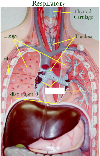
FREE Human Body Systems Labeling with Answer Sheets The free skeletal system labeling sheet includes a fill-in-the-blanks labeling of the main bones on the body. The free respiratory system labeling sheet includes a blank diagram to fill in the trachea, bronchi, lungs, and larynx. The free nervous system labeling sheet includes blanks to label parts of the brain, spinal cord, ganglion, and nerves.

labeled brain anatomy - Clip Art Library
14 Informative Facts, Diagram & Parts Of Human Brain For Kids Human brain and the spinal cord are covered by three layers of tissue collectively known as the meninges. A clear fluid called cerebrospinal fluid flows between these layers (10) (11). The left part of the brain is responsible for analytical thinking. The right part helps in creative thinking (12).
Brain Viewed from Above | ClipArt ETC
Draw And Label The Human Brain Structure - Draw Neat ... The brain structure is composed of three main parts: The structure and function of the human brain. The skull is composed of two sets of bones cranial and facial. Brain diagram, pituitary gland, anatomy, adhd, draw, search, to draw. How to draw a the human brain easy and step by step.
Print Activity 5: Examining the Human Torso Model flashcards | Easy Notecards
Label-free imaging of human brain tissue at subcellular ... The human neocortex is conventionally divided into six layers [ 28 ]. These layers - molecular, outer granular, outer pyramidal, inner granular, inner pyramidal, and pleomorphic layers - are roughly distinguishable in MPM data ( Figure 2 A; representative higher-magnification images from each layer are shown in Figure S3 ).
Post a Comment for "44 labels of the human brain"