41 chlamydomonas diagram with labels
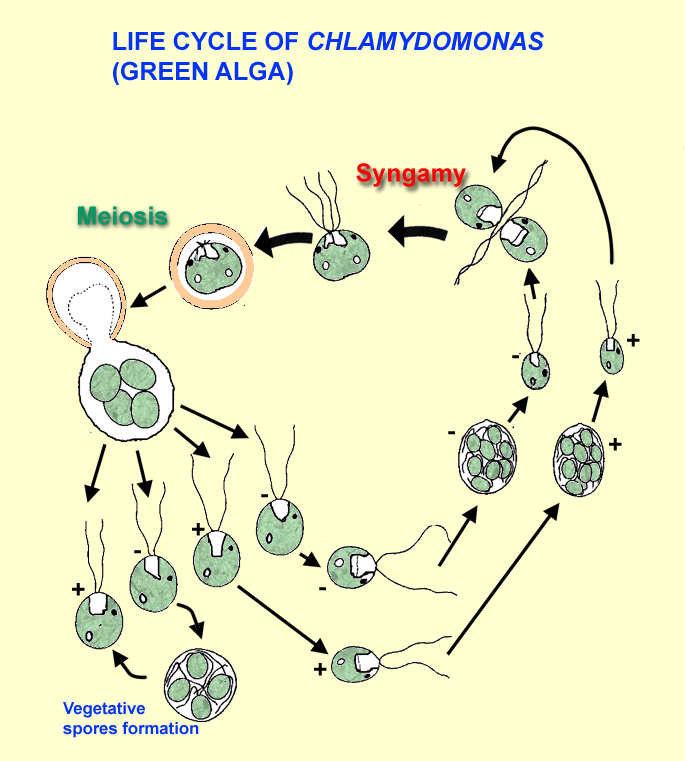
Draw a labelled diagram of Chlamydomonas. - Brainly.in 6 Oct 2019 — This is Expert Verified Answer · Chlamydomonas is a unicellular, motile freshwater species belonging to the genus of green algae. · They are oval, ... Life Cycle of Chlamydomonas (With Diagram) - Biology Discussion Life Cycle of Chlamydomonas (With Diagram) Article Shared by ADVERTISEMENTS: In this article we will discuss about the asexual and sexual methods of reproduction that occur in the life cycle of chlamydomonas. 1. Asexual Reproduction: It takes place by following methods:
Substancial | PDF | United Kingdom | Spain - Scribd substancial - Free ebook download as Text File (.txt), PDF File (.pdf) or read book online for free. contains some random words for machine learning natural language processing

Chlamydomonas diagram with labels
Higher Education Support | McGraw Hill Higher Education Learn more about McGraw-Hill products and services, get support, request permissions, and more. MIT - Massachusetts Institute of Technology a aa aaa aaaa aaacn aaah aaai aaas aab aabb aac aacc aace aachen aacom aacs aacsb aad aadvantage aae aaf aafp aag aah aai aaj aal aalborg aalib aaliyah aall aalto aam ... Biological drawings. Structure of Chlamydomonas. Learning Resources for ... Structure of Chlamydomonas: Next Drawing > Chlamydomonas is the name given to a genus of microscopic, unicellular green plants (algae) which live in fresh water. Typically their single-cell body is approximately spherical, about 0.02 mm across, with a cell wall surrounding the cytoplasm and a central nucleus.
Chlamydomonas diagram with labels. Amoeba Diagram Pictures, Images and Stock Photos Browse 116 amoeba diagram stock photos and images available, or start a new search to explore more stock photos and images. Anatomy of an amoeba. Amoeba unicellular animal with pseudopods that lives in fresh or saltwater. Anatomy of an amoeba. Vector illustration for medical, educational and science use. Amoeba labeled vector illustration. Draw a neat labelled diagram. Chlamydomonas - Biology Diagram Draw a neat labelled diagram. Chlamydomonas Advertisement Remove all ads Solution Concept: Salient Features of Major Plant Groups Under Cryptogams Report Error Is there an error in this question or solution? Advertisement Remove all ads Chapter 3: Kingdom Plantae - Exercise [Page 28] Q 8. (B) Q 8. (A) Q 8. (C) APPEARS IN Lifestyle | Daily Life | News | The Sydney Morning Herald The latest Lifestyle | Daily Life news, tips, opinion and advice from The Sydney Morning Herald covering life and relationships, beauty, fashion, health & wellbeing Use this labeled diagram of a chlamydomonas cell to - Course Hero Use this labeled diagram of a Chlamydomonas cell to address the following two questions. 32. Which of the following statements correctly identifies aspects related to photosynthesis and/or respiration? 1. Acetyl CoA is most often found in G. 2. NADPH accumulates in C. 3. ATP is found in F. 4.
Structure of Chlamydomonas (With Diagram) | Chlorophyta In this article we will discuss about the structure of chlamydomonas with the help of suitable diagrams. Chlamydomonas is unicellular, motile green algae. The thallus is represented by a single cell. It is about 20 p,-30|i in length and 20 µ in diameter. The shape of thallus can be oval, spherical, oblong, ellipsoidal or pyriform. Morphology of Chlamydomonas (With Diagram) | Algae - Biology Discussion In this article we will discuss about the external morphology of chlamydomonas. Also learn about its Neuromotor Apparatus, Electron Micrograph, Palmella-Stage with suitable diagram. 1. The organism is an unicellular alga (Fig. 11). 2. The thallus is spherical to oblong in shape but some species are pyriform or ovoid. ADVERTISEMENTS: 3. Characterization techniques for nanoparticles: comparison and ... The size distribution of their particles depended on the pH of the culture medium. The Ag NP toxicity on the green alga Chlamydomonas acidophila was pH-dependent as shown by the cytotoxicity mediated through the induction of oxidative stress. 227. Pavlopoulou et al. monitored the synthesis of Pt NPs using pH-responsive microgel particles. SAXS ... LABORATORY 9 - Susquehanna University CHLOROPHYTA & CHAROPHYTA This laboratory will cover those taxa that collectively are called the Green Algae. Like other members of the Viridiplantae, they tend to have walls of cellulose, chloroplasts with chlorophylls a&b, store starch as a photosynthate, and, when they make motile cells, have two anteriorly-directed whiplash flagella.
Chlamydomonas reinhardtii - an overview | ScienceDirect Topics Chlamydomonas reinhardtii cells are oval shaped, c. 10 μm in length and 3 μm in width, with two flagellae at their anterior end ( Figure 1 ). The cells contain a single chloroplast occupying 40% of the cell volume and several mitochondria. These cells exist as mating-type (+) or mating-type (-). Structure and Diagram of Volvox and Their Functions - NotesHippo Volvox Structure: Diagram of Volvox with Label The cells of anterior end possess bigger eye spots than those of posterior end cells. The cells of posterior side become reproductive on maturity. Thus, spherical or round colony of Volvox shows clear polarity. Cell structure of volvox colony are Chlamydomonas type. Modular, cascade-like transcriptional program of regeneration ... Aug 04, 2022 · In Chlamydomonas, radial spoke protein synthesis reaches its maximum rate 30–60 min after the flagella have begun assembling onto pre-existing basal bodies (Remillard and Witman, 1982). This timing roughly matches the delay of 1 hr seen in our data between the peak expression of genes involved in ciliary assembly (cluster 2) and genes ... Structure of Chlamydomonas (With Diagram) | Genetics - Biology Discussion In this article we will discuss about the structure of chlamydomonas (explained with labelled diagram). The unicellular green alga Chlamydomonas is haploid with a single nucleus, a chloroplast and several mitochondria (Fig. 9.3). It can reproduce asexually as well as sexually by fusion of gametes of opposite mating types (mt + and mt - ).

Cladocera, water fleas: taxonomy, diversity, anatomy, drawings at GeoChemBio
Chlamydomonas: Position, Occurrence and Structure (With Diagrams) Chlamydomonas is unicellular, motile green algae. The thallus is represented by a single cell. It is about 20 p,-30|i in length and 20 µ in diameter. The shape of thallus can be oval, spherical, oblong, ellipsoidal or pyriform. The pyriform or pear shaped thalli are common, they have narrow anterior end and a broad posterior end (Fig. 1).

LABORATORY 20
Chloroplast Structure and Function in detail with Labelled Diagram The chloroplasts are the cell organelles which consist of these pigments. The 3 types of pigments present in plants are chlorophyll, carotenoids, and anthocyanins. Chlorophyll imparts the green color to plants. Plastids are membrane-bound cytoplasmic organelles that can be found in the cells of plants and algae.

Antibody producing algae | MMG 233 2013 Genetics & Genomics Wiki | FANDOM powered by Wikia
Structure of Volvox (With Diagram) | Chlorophyta - Biology Discussion The cells of Volvox colony are Chlamydomonas type. Every cell has its own mucilage sheath (Fig. 1 B). The mucilage envelope of colony appears angular due to compression between cells. The cells are connected to each other through cytoplasmic strands. In some species of Volvox the cytoplasmic connections or strands are not present.

Chlamydomonas | ClipArt ETC
Chlamydomonas - Meaning, Structure, Life Cycle, Function and FAQs - VEDANTU Chlamydomonas structure is a single cell used to represent the thallus. It measures approximately 20 p,-30|i in length and 20 p,-30|i in diameter. Thallus may be circular, rectangular, oblong, ellipsoidal, or pyriform in shape. The pyriform or pear-shaped thalli, which have a narrow anterior end and a wide posterior end, are normal.
[Groenwieren: Green algae: Chlorophytae
Microorganisms: Friend and Foe Class 8 Extra Questions ... Oct 11, 2019 · Pull out a gram or bean plant from the field. Observe its roots. You will find round struc¬tures called root nodules on the roots. Draw a diagram of the root and show the root nod¬ules. Answer: Question 2. Collect the labels from the bottles of jams and jellie on the labels. Answer: Do it yourself. Question 3. Visit a dcotor.

Cianna Gibson Science 10 Ms. Ward: Micro-organism Lab
Amoeba Diagram Illustrations, Royalty-Free Vector Graphics ... - iStock Browse 65 amoeba diagram stock illustrations and vector graphics available royalty-free, or start a new search to explore more great stock images and vector art. Newest results. Anatomy of an amoeba. Amoeba unicellular animal with pseudopods that lives in fresh or saltwater. Anatomy of an amoeba.

Chlamydomonas – Parts Labeled | Radesaal the science group
Biological drawings. Structure of Chlamydomonas. Learning ... Dec 27, 2012 - Biological drawings of Protista, Structure of Chlamydomonas, Resources for Biology Education by D G Mackean.

Chlamydomonas anatomy. It's a unicellular organism, yet it's got a way to make it's energy into ...
Chlamydomonas - Wikipedia Drawings of Chlamydomonas caudata Wille. [1] Cross section of a Chlamydomonas reinhardtii cell Light micrograph of Chlamydomonas with two flagella just visible at bottom left Chlamydomonas globosa, again with two flagella just visible at bottom left

draw labelled diagram of chlamydomonas.answer me fast if you want brainliest first comer first ...
Clear Labeled Diagram Of Volvox - A Rubisco Binding Protein Is Required ... 12.10.2021 · labeled in the chlamydomonas diagram. Mean that a chlamydomonas is primitive itself. Volvox, chlamydomonas, and the evolution of multicellularity. The mucilage envelope of colony appears angular due to compression between cells. The cells are connected to each other through cytoplasmic strands.

Comparison of ascorbate peroxidase amino acid sequences. A: Amino acid... | Download Scientific ...
Describe the structure of chlamydomonas with neat labelled diagram ... answeredOct 30, 2020by Naaji(56.8kpoints) selectedOct 30, 2020by Jaini Best answer 1. Chlamydomonas is a simple, unicellular, motile fresh water algae. They are oval, spherical or pyriform in shape. 2. The cell is surrounded by a thin and firm cell wall made of cellulose. 3. The cytoplasm is seen in between the cell membrane and the chloroplast. 4.

Sample Descriptive Lab Report
Eye Diagram With Labels and detailed description - BYJUS A brief description of the eye along with a well-labelled diagram is given below for reference. Well-Labelled Diagram of Eye The anterior chamber of the eye is the space between the cornea and the iris and is filled with a lubricating fluid, aqueous humour. The vascular layer of the eye, known as the choroid contains the connective tissue.
Cells | Special Issue : Chlamydomonas Cell Biology
Biological drawings. Structure of Chlamydomonas. Learning Resources for ... Structure of Chlamydomonas: Next Drawing > Chlamydomonas is the name given to a genus of microscopic, unicellular green plants (algae) which live in fresh water. Typically their single-cell body is approximately spherical, about 0.02 mm across, with a cell wall surrounding the cytoplasm and a central nucleus.

Contoh Explanation Text Photosynthesis - 6 Contoh x
MIT - Massachusetts Institute of Technology a aa aaa aaaa aaacn aaah aaai aaas aab aabb aac aacc aace aachen aacom aacs aacsb aad aadvantage aae aaf aafp aag aah aai aaj aal aalborg aalib aaliyah aall aalto aam ...

35 Can You Label A Diagram Of The Alternation Of Generations Life Cycle_ - Labels Database 2020
Higher Education Support | McGraw Hill Higher Education Learn more about McGraw-Hill products and services, get support, request permissions, and more.
Root Canal Treatment Dental Infographic All Stock Vector 402395899 - Shutterstock

The Chlamydomonas Genome Reveals the Evolution of Key Animal and Plant Functions | Science
Post a Comment for "41 chlamydomonas diagram with labels"